PhD. Student - Jihan M. Zoghbi
Presentation
|
Possui graduação em Ciência da Computação pela Universidade Presbiteriana Mackenzie (2002) mestrado em Ciência da Computação pela Universidade de São Paulo (2011). Tem experiência na área de Ciência da Computação, com ênfase em Computação Gráfica e processamento de imagens médicas. |
CONTACT
This email address is being protected from spambots. You need JavaScript enabled to view it.
|
 Projects
Applying Geometric Deformable Models to Segment Brain Lesions from Multi-spectral MR Images
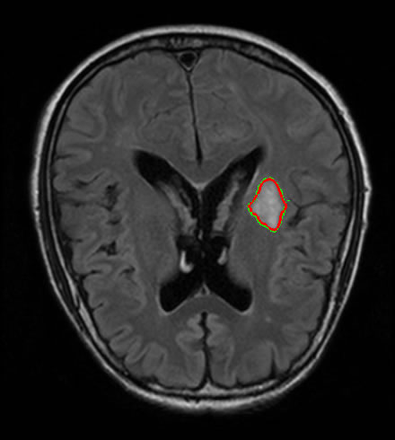
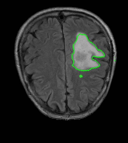
The main objective of medical image segmentation is the partition of the image into several homogeneous regions with respect to one or more characteristics according to the diagnostic needs. The segmentation of brain lesion helps doctor to see the abnormal areas and their boundaries, in order to assist in the diagnosis of disease and provide follow-up. Furthermore, there are certain cases where the clinical diagnosis of these images discards the possibility of performing a biopsy in the patient.
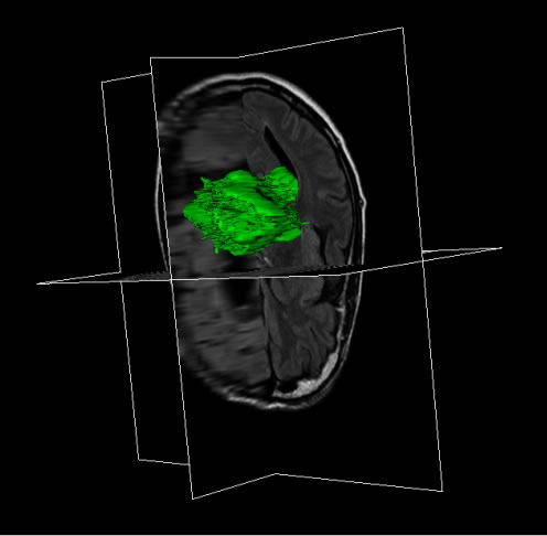
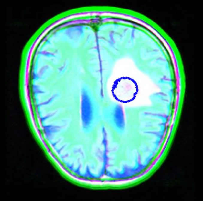
Currently, magnetic resonance imaging (MRI) is considered the most efficient for the diagnosis of brain lesions, although there are other methods of complementary image. MRI modality can provide high spatial resolution images of anatomical soft tissue in different contrasts: FLAIR (Fluid attenuated inversion recovery), T1and T2- weighted images. Due to their facility to adapt to topological change, deformable model are widely used in the literature to segment brain lesions using one type of contrasts. In this paper we applied and compare two methods using deformable models: Mumford-Shah model via level set (MS) and Geodesic Active Contour (GAC) models, in order to segment multi-spectral volumetric brain magnetic resonance (MR) images that combines FLAIR, T1 and T2- weighted images. Results showed that segmentation using multi-spectral images provide superior results than using each contrast alone. An extension of MS model to 3D has been elaborated to segment multi-spectral images.
As a part of this work, a free open-source software has been developed and integrated to platform MedSquare (http://sourceforge.net/projects/medsquare/branches/levelset) to visualize and segment brain lesions.